TraCS Pilot Award recipient continues to pioneer X-ray technology
In 2016, UNC researcher Otto Zhou received NC TraCS funding with an Improving Human Health pilot award for: A low-dose and high-sensitivity 3D intraoral imaging device for improving human health, which helped fund a clinical evaluation that compared the new 3D device to standard radiography. Now, with the launch of a new company in RTP, Zhou and fellow researcher Jianping Lu hope to have the new device in dentist's hands by the end of 2021.
From Endeavors:
A Sharper Image
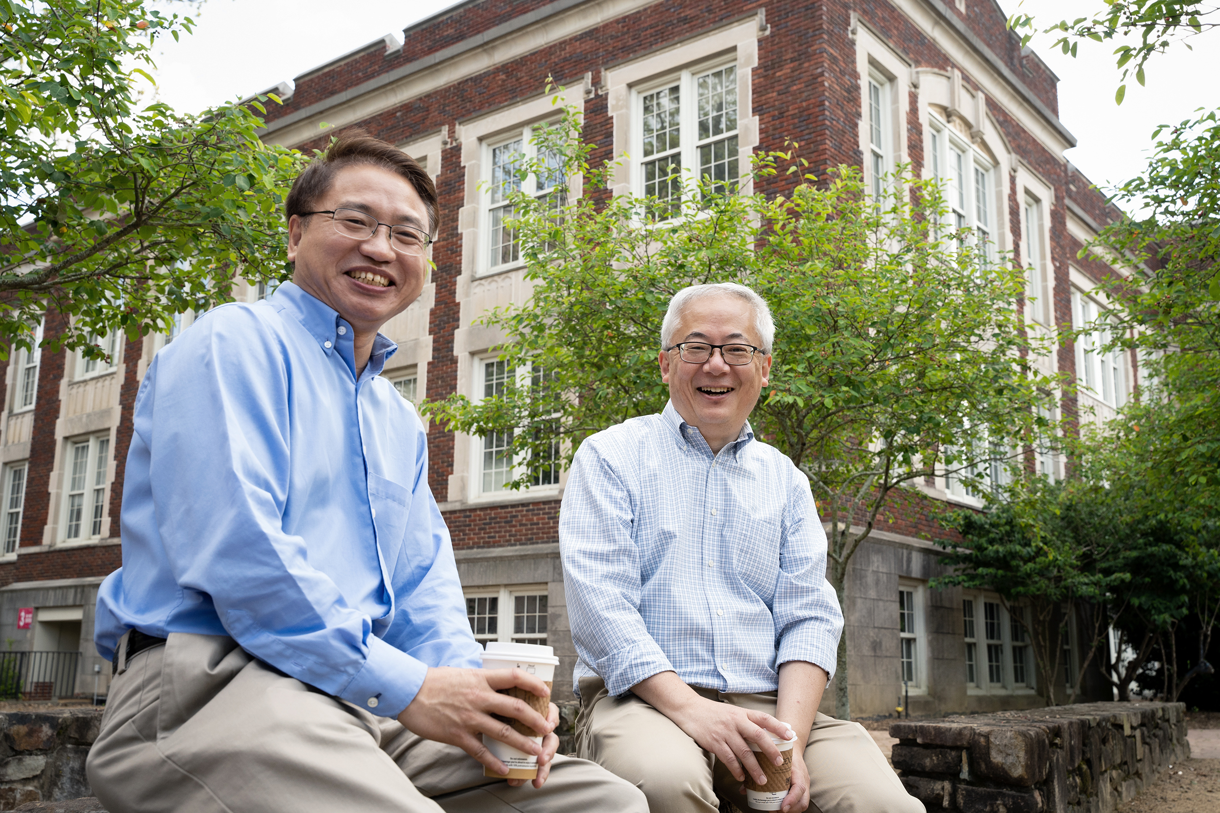
UNC physics professors Jianping Lu and Otto Zhou have been working together since the early 2000s, using atom-sized devices called carbon nanotubes to improve X-ray technology. (photo by Elise Mahon)
UNC researchers Jianping Lu and Otto Zhou have spent the last two decades refining technology that makes X-ray machines smaller, faster, safer, and sharper — research that's changing the world of dentistry, medicine, and security.
In 2005, UNC physicist Jianping Lu noticed that his 5-year-old son was having trouble breathing. He took him to a local doctor, who ordered a CT scan to see what might be happening inside the boy's lungs. The resulting picture wasn't clear enough for the doctor to give them a diagnosis, and because they'd discussed a potential — and risky — surgery to fix the problem, Lu got a second opinion.
"The second CT scan was slightly better," he remembers. "The doctor said maybe we should wait a couple years, and he may grow out of it."
Luckily, that's exactly what happened and his son's condition improved. But the experience showed Lu why the research he was doing at the time with UNC materials scientist Otto Zhou was so important.
Since the early 2000s, Lu and Zhou have been collaborating to improve X-ray technology in the medical field. At the time, both were working with carbon nanotubes — tiny tubes made of a single layer of carbon atoms. Lu was theorizing how they might transmit electrons in a simpler, less resistive way, and Zhou was testing the application of that theory in his lab. Their experiments lined up.
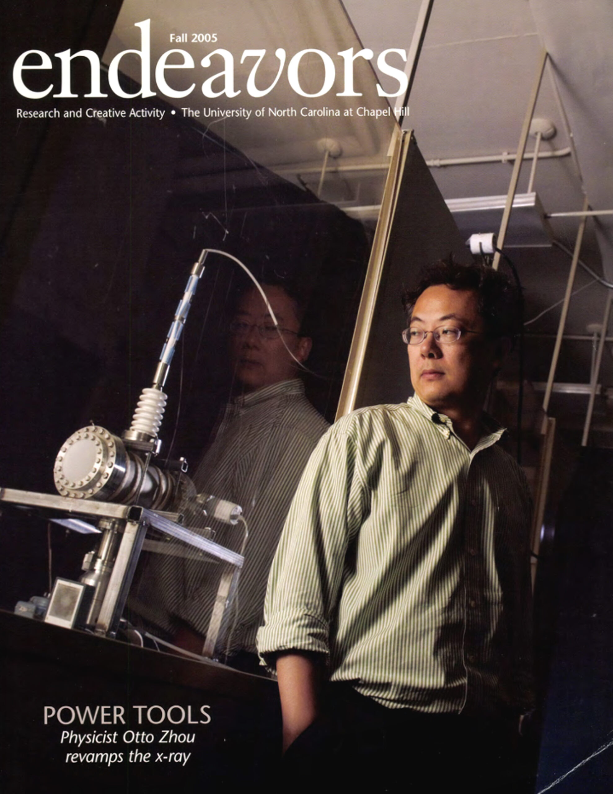
Zhou was featured on the Fall 2005 cover of Endeavors for his research.
Since then, Lu and Zhou have worked on numerous projects across campus to create better X-ray sources and imaging systems in the dental and medical fields.
"Everything we work on requires a close team of people from different disciplines bringing their expertise together," Zhou says. "We have engineers, physicists, radiologists, physicians. That really helps us move forward."
Endeavors featured this research and placed Zhou on the cover of the Fall 2005 issue. The article discussed their early projects, creation of a spinoff company called Xintek, and the prototypes they were able to produce. But it takes a long time for technology like this to come to the commercial market.
"There's a lot of engineering fine-tuning that needs to happen," Zhou says. "It's a challenging, large effort that requires a lot of persistence."
Sixteen years later, one of Zhou and Lu's projects will finally be available to doctors: a 3D dental X-ray device, which will provide a big upgrade to current 2D technology. And that's just one of many projects coming down the pike.
Smaller, faster, clearer
An X-ray at the dentist's office used to involve going into a separate room where a dental hygienist would place a heavy lead vest over your shoulders and aim the tube of a giant machine at one side of your mouth. They'd leave the room, press a button, and take the X-ray. They'd do this a few times, coming back in to move the tube each time, eventually getting a full picture of the mouth. The whole process would take about 10 minutes.
Now when you visit the dentist, you stay in the same room your cleaning is conducted in. The dental hygienist places a small device in your mouth and asks you to bite down. Within a few seconds, she's captured 2-dimensional images of all your teeth.
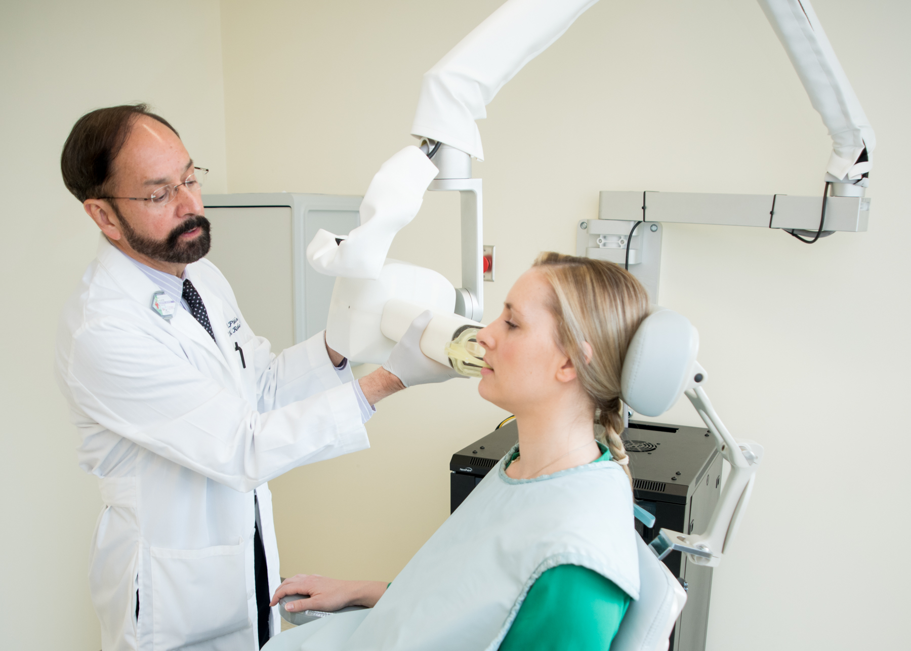
Enrique Platin, a now retired UNC Adams School of Dentistry professor, uses a 2D X-ray machine on a patient. Platin is a collaborator on Zhou and Lu’s 3D dental X-ray project. (photo courtesy of the UNC Adams School of Dentistry)
This is done using a conventional X-ray source, which produces electrons and uses high voltage to accelerate them, forcing them against a Tungsten target that generates enough radiation to produce an image. More than 99 percent of the energy used to produce the X-ray turns into heat — which means less than 1 percent makes the image. It takes a long time to cool down and wastes a lot of energy.
"It's similar to a space heater," Zhou says. "You apply a current to a piece of metal and it becomes really hot — about 1000oC."
That amount of heat eventually causes the metal filaments within the machine to burn out — and they cost about $6,000 to replace.
Zhou and Lu's X-ray device uses carbon nanotubes, which are basically sharp, atom-sized needles. These require a lot less energy to work, removing the need for the large heating component a traditional X-ray machine requires.
"It's sort of like an LED light that can be flipped on and off continuously," Zhou says.
Lower voltage means smaller devices and a faster, clearer image. It also means a more efficient machine with less upkeep and a wide range of applications. Simply put, it's cheaper and more versatile.
Read the full article at endeavors.unc.edu.
Dr. Zhou has also received TraCS funding for the following:
TraCS $50K Pilot Award
April 2009
Nanotechnology Enabled Compact Microbeam Radiation Therapy System for Cancer Treatment and Research
FastTraCS (commercialization) Grant
August 2018
Development of a compact, low-dose and low-cost 3D orthopedic imaging device