Liquid Metal 'Nano-Terminators' Target Cancer Cells
The new cancer drug delivery system improved efficacy of standard chemotherapy for ovarian cancer in mice with limited toxicity.
RALEIGH, NC – Researchers at North Carolina State University and the UNC School of Medicine have developed a new drug delivery technique that uses a biodegradable liquid metal to target cancer cells while limiting toxicity to normal cells. The development of this novel method, published in Nature Communications, may enhance the effectiveness of cancer drugs already in use and help doctors locate tumors.
“We can produce these molecules in bulk, and they appear to be wholly biodegradable with very low toxicity,” says Zhen Gu, PhD, senior author of a Nature Communications paper and assistant professor in the joint biomedical engineering program at NC State and UNC. His team has developed other nano-sized drug delivery systems. “One of the advantages of this technique is that these liquid metal drug carriers – or ‘nano-terminators’ – are very easy to make,” he said.
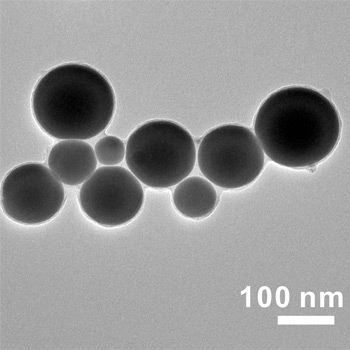
To create the nano-terminators, researchers placed the bulk liquid metal (gallium indium alloy) into a solution that contained two types of molecules called polymeric ligands. The solution was then hit with ultrasound, which forced the liquid metal to burst into nanoscale droplets approximately 100 nanometers in diameter. The ligands in the solution attached to the surface of the droplets as they broke away from the bulk liquid metal. Meanwhile, an oxidized “skin” forms on the surface of the nanodroplets. The oxidized skin, together with the ligands, prevents the nanodroplets from fusing back together.
The anticancer drug doxorubicin (Dox) was then introduced into the solution. One of the ligands on the nanodroplet sucked up the Dox and held on to it. These drug-laden nanodroplets could then be separated from the solution and introduced into the bloodstream.
The second type of ligand on the nanodroplets effectively sought out cancer cells, causing receptors on the surface of the cancer cell to latch on to the nanodroplets. The cancer cell then absorbed the nanodroplets.
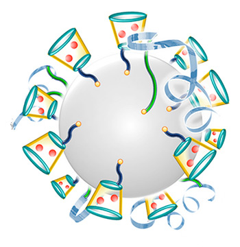
Once absorbed, the higher level of acidity inside the cancer cell dissolved the oxidized skin of the nanodroplets. This released the ligands, which then went on to release the Dox inside the cell.
"Without the oxidized skin and ligands, the nanodroplets fuse together, forming larger drops of liquid metal," says Michael Dickey, a co-author on the paper and professor in of chemical and biomolecular engineering at NC State. "These larger droplets are fairly easy to detect using diagnostic techniques, which can potentially help doctors locate tumors."
As the liquid metal continued to react with the acidic environment in the cancer cell and dissolve, gallium ions were released. Interestingly, these gallium ions enhanced the performance of Dox – including the drug’s effectiveness against drug-resistant cell lines.
In addition, this process gradually degraded the metal, minimizing long-term toxicity.
"Based on in vitro tests, we believe the liquid metal degrades completely in a matter of days into a form that the body can successfully absorb or filter out, without notable toxic effects," says Yue Lu, a graduate student in Gu’s lab and author on the Nature Communications paper.
The researchers have tested the liquid metal technique in a mouse model, and found that it is significantly more effective than Dox alone at inhibiting the growth of ovarian cancer tumors. Importantly, the researchers tracked the mice for up to 90 days, and found no signs of toxicity related to the liquid metal.
"This was a proof-of-concept study, but very encouraging," Gu says. "This drug carrier is transformable: smashed from bulk material, fused inside cancer cells, and eventually degraded and cleared. We are hoping to do additional testing in a large animal study to get closer to potential clinical trials."
This work was supported by the NC TraCS Institute, NIH’s Clinical and Translational Science Awards at UNC-CH, and by the National Science Foundation.
Article originally posted at UNC Health Care Newsroom
Media Contacts:
Matt Shipman, 919-515-6386, This email address is being protected from spambots. You need JavaScript enabled to view it.;
Mark Derewicz, 919-923-0959, This email address is being protected from spambots. You need JavaScript enabled to view it.